
Keratoacanthoma: Symptoms, Causes, and Prevention by Dr Yeung Ho Hong 楊浩康
What is Keratoacanthoma: Causes, Clinical Features, and Treatment Strategies
Keratoacanthoma (角化棘皮瘤) is a rapidly growing skin tumor with local destructive potential, primarily originating from the infundibular portion of the hair follicle. Although its exact pathogenesis is not fully understood, it is currently believed to be associated with abnormal regulation of the WNT signaling pathway and mutations in the tumor suppressor gene TP53. These molecular changes contribute to the rapid growth characteristic of keratoacanthoma, often forming noticeable nodules within a short period.
Risk factors for keratoacanthoma include prolonged ultraviolet (UV) exposure, contact with chemical carcinogens, skin trauma (such as surgery or radiation), and human papillomavirus infection. These external and internal factors may trigger abnormal proliferation of skin cells, leading to the development of keratoacanthoma. Since this type of tumor frequently appears on sun-exposed areas such as the face and upper limbs, sun protection is particularly important during summer or outdoor activities.
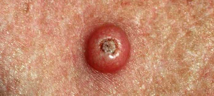
Clinically, keratoacanthoma typically presents as a solitary, rapidly enlarging nodule with well-defined borders, a firm texture, and a red or flesh-colored appearance. A prominent keratin plug is often observed at the center of the lesion, and upon removal, the lesion takes on a characteristic “volcano crater” appearance. While its appearance may resemble other skin conditions such as squamous cell carcinoma, basal cell carcinoma, or seborrheic keratosis, most cases of keratoacanthoma are benign. To ensure diagnostic accuracy, histopathological examination is often required to differentiate it from these other lesions and avoid misdiagnosis as a malignant tumor.
Additionally, some keratoacanthomas may spontaneously regress within 4 to 6 months, potentially leaving a scar. However, since its clinical progression is unpredictable, surgical excision remains the most common and safest treatment approach. Complete surgical removal not only eliminates the lesion but also provides tissue for pathological analysis to rule out the possibility of well-differentiated squamous cell carcinoma. For most patients, the prognosis following surgery is excellent, with smooth wound healing and minimal impact on quality of life.
In clinical practice, many patients and their families harbor common misconceptions about keratoacanthoma. Some believe that all rapidly growing skin tumors are malignant, leading to excessive fear and inappropriate treatment. Others mistakenly assume that all keratoacanthomas will resolve on their own without intervention. In reality, while some lesions may be self-limiting, their course is unpredictable, and there is a risk of confusion with squamous cell carcinoma. Thus, understanding its pathological features and undergoing timely medical evaluation are crucial.
In summary, keratoacanthoma (角化棘皮瘤) is a rapidly growing, locally destructive benign skin tumor that resembles malignant lesions, necessitating careful diagnosis. Through surgical excision, histopathological examination, and appropriate adjuvant therapy, most patients achieve favorable outcomes and prognosis. Daily sun protection, avoidance of chemical and physical irritants, and regular skin checkups are key preventive measures. Accurate diagnosis and proactive treatment strategies not only alleviate patient concerns but also safeguard skin health and enhance quality of life.